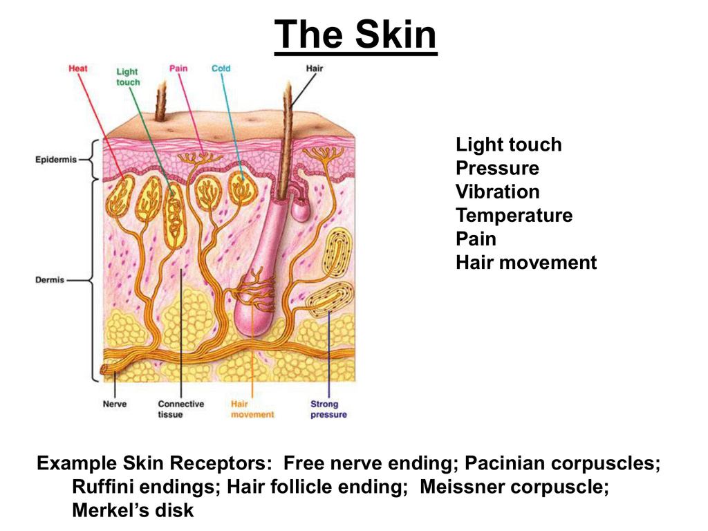
The above description corresponds to the classical structure of a Ruffini corpuscle. The axon loses the myelin sheath and bifurcates in two before encapsulating to form branched nerve endings. In this capsule, the nerve endings are anchored between collagen fibers of connective tissue.
#Ruffini endings free#
They are formed by numerous free nerve endings, originating from a common myelinated axon, which are encapsulated forming a cylindrical structure. All this because they adjust to the new surroundings.įinally, Ruffini's corpuscles found in the joint capsules of birds and mammals, are located only in the areas that are inside the fibrous layer and the ligaments of the capsule. It can be assumed that the structural change in the connective tissue (injuries, bad position of the joints, scars, degenerative processes, aging) also leads to a change in the corpuscles of Ruffini. These corpuscles are quite small in size and are not very numerous. Given their ability to detect signals with very small receptive fields, Ruffini endings fall within the classification of type I mechanoreceptors. In addition to detecting these types of static stimuli, they also respond to dynamic factors such as joint angle, stimulus speed, and stretch. Slowly adapting mechanoreceptors are capable of detecting sustained or prolonged pressure stimuli on the skin, as well as slight deformations produced by stretching it. Additionally, they are capable of perceiving low levels of mechanical deformation of the skin, even in the deepest layers of the skin. They are cutaneous sensory receptors, that is, located in the skin, being specialized in perceiving temperature variations above or below body temperature. However, all of them are mechanoreceptors that adapt slowly to stimulus and perceive stimuli in small receptive fields. The Ruffini corpuscles found in each of the above locations show slight variations in structure.
#Ruffini endings skin#
They are located both in the dermis and in the hypodermis of the glabrous and hairy skin of mammals and marsupials, as well as in the menisci, ligaments and joint capsules of the joints of some birds and mammals. These receivers are named after the Italian physician and biologist Angelo Ruffini (1864-1929). This capsule can be composed of collagen synthesized by fibroblasts or perineural cells. These consist of a single myelinated axon that branches into multiple nerve endings that anchor inside a capsule. The Ruffini corpuscles They are sensory receptors that respond to mechanical stimuli and subtle variations in temperature. Classification of mechanoreceptors based on their function.1639051519).Video: Meissner corpuscle, Pacinian corpuscle, Ruffini ending, Merkel disc Content Conclusions: These findings indicate that NT-4/5 is required for the regeneration of periodontal Ruffini endings after transection of the IAN. However, the neural density in the NT-4/5 homozygous mice never recovered to the control level at postoperative 28 days when the periodontal Ruffini endings in the wild-type mice showed the same terminal morphology and neural density. Quantitative analysis demonstrated a constant lower neural density in the restricted area at each stage (>15% reduction) throughout experimental stages. They re-appeared in both animals at postoperative 7 days, and their regeneration has proceeded later with increasing of neural density in both types. At 3 days after nerve injury, the PGP 9.5-positive neural elements had completely disappeared in both types. We failed to find such typical Ruffini endings in the periodontal ligament of the homozygous mice: A majority of the periodontal Ruffini endings remained to show smooth outlines. Results: In control group, without nerve injury, immunohistochemistry for PGP 9.5 demonstrated the presence of immature Ruffini endings in the periodontal ligament of the NT-4/5 (-/-) homozygous mice. The chronological neural density was calculated with a computer-assisted image analyzer.

The animals whose inferior alveolar nerve (IAN) was cut were processed for immunohistochemistry for protein gene product 9.5 (PGP 9.5), a general neuronal marker. Methods: NT-4/5 homozygous and wild-type mice were used in this study. Our recent studies using BDNF-deficient mice demonstrated a delayed in regeneration of the periodontal Ruffini endings, and furthermore suggested the involvement of neurotrophin-4/5 (NT-4/5) in their regeneration. Objectives: The expression of TrkB, a high affinity neurotrophin receptors, have been shown in the periodontal Ruffini endings, suggesting the involvement of brain-derived neurotrophin (BDNF), a ligand for TrkB, in the development and regeneration of this mechanoreceptor.


 0 kommentar(er)
